|
ADVANCED IMAGING CENTER PHYSICIAN NEWS |
October 18, 1999 |
Advanced Topics in Neuroradiology: MR DIFFUSION and PERFUSION
|
ADVANCED IMAGING CENTER PHYSICIAN NEWS |
October 18, 1999 |
Q. What is MR Diffusion imaging?
MR diffusion imaging is a non-invasive fast MRI technique that evaluates molecular diffusion in the tissue.
Q. What MRI techniques are utilized in Diffusion imaging?
Ultrafast techniques such as Echoplanar Imaging (EPI) or HAlf-Fourier Single-shot Turbo Spin Echo (HASTE). High-performance gradients are needed. The study is performed in a few seconds.
Q. What are some applications?
In the brain, it is used to determine hyperacute and acute strokes (detectable from minutes to days). Conventional imaging may not be able to detect hyperacute strokes or differentiate acute from chronic infarcts. With MR diffusion, detection of acute infarcts is immediate and differentiation from old infarcts is obvious.
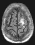
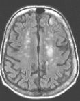
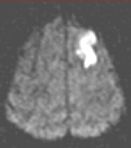
Routine FLAIR MRI images (left) cannot differentiate between acute infarcts and chronic ischemic changes/infarcts. The diffusion MRI (right) clearly demonstrates an acute infarct in the left frontoparietal region (the chronic changes do not "light up" on the diffusion study).
Q. Any other applications besides cranial infarcts?
Recently, diffusion has been used to effectively differentiate between acute traumatic vs. pathologic compression fractures in the spine. This is exciting since no other diagnostic test, short of a biopsy, can make the differentiation.
Q. Is Diffusion imaging available at AIC?
Yes! It has been available and routinely used since January 1999 on the high-field 1.5 Tesla short-bore Siemens Symphony MRI. The ultra high gradients of this system allow for ultrafast imaging including diffusion. Hospital patients have been transported to AIC for this test by ambulance.
Q. I have heard of MR Perfusion imaging. Is that also available at AIC?
It will be available shortly (in October 1999).
Q. How is MR Perfusion different from MR Diffusion?
MR Perfusion (with the use of Gadolinium contrast) determines areas of decreased perfusion in the brain (ischemic areas). Comparison of perfusion and diffusion images enables one to determine what portion of the CVA is reversible (i.e., reversible ischemia vs. non-reversible infarct).
For more information, please call me personally at (661) 949-8111.
Ray Hashemi, MD, PhD,
Director