 |
ADVANCED IMAGING CENTER PHYSICIAN NEWS |
September 15, 2003 |
DIAGNOSIS OF SUBARACHNOID HEMORRHAGE (SAH)
with FLAIR MRI IMAGING
 |
 |
 |
 |
 |
| Fig.1 | Fig.2a | Fig.2b | Fig.3a | Fig.3b |
|
ADVANCED IMAGING CENTER PHYSICIAN NEWS |
September 15, 2003 |
|
|
|
|
|
| Fig.1 | Fig.2a | Fig.2b | Fig.3a | Fig.3b |
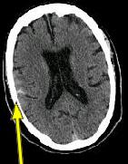
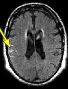
CLINICAL HISTORY: This is a 70-year-old male who presented with headaches and dizziness to the office of Dr. Pamela DeSilva* after a fall. He was referred to AIC for a head CT.
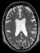
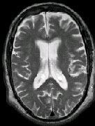
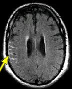
IMAGING FINDINGS: The CT was performed on AIC’s new 16-slice helical multi-slice CT (MSCT). Both routine single-slice and thin-section helical scans were obtained and 3D/Multiplanar images were performed on the Advanced 3D workstation. Fig. 1 shows an image at the level of the lateral ventricles with possible high density within the sulci on the right (arrow). To confirm this finding, an MRI with contrast was then obtained on AIC’s 1.5-Tesla high-field, short-bore Siemens MRI. Fig. 2a-b are T2-weighted images at the same level showing NO definite abnormality. T1-weighted images (not shown) were also negative. The post-contrast images (not shown) showed NO abnormal enhancement in this region, either. Diffusion-weighted images were also negative. However, Fig. 3a-b, which are FLAIR images, demonstrate clear hyperintensity within multiple sulci involving the right temporo-parietal area (arrows).
DIAGNOSIS: The FLAIR images are compatible with Subarachnoid disease. The Differential Diagnosis includes: (1) Subarachnoid Hemorrhage (SAH); (2) Meningitis; (3) Infarct; (4) Flow Artifact. Lack of enhancement is against meningitis. Acute infarct was ruled out on the basis of negative Diffusion MRI and lack of cytotoxic edema on the FLAIR images. Focal abnormality is against flow artifact (which is more diffuse and usually in the posterior fossa on FLAIR images). The clinical history of trauma and the high-density on CT and hyperintensity on FLAIR images within the sulci are diagnostic of Acute Post-traumatic Subarachnoid Hemorrhage (SAH).
DISCUSSION: MRI FLAIR imaging is the modality of choice for diagnosis of supratentorial SAH. In the posterior fossa, however, CT is superior due to flow artifacts on FLAIR images in the peripontine/ambient cisterns. A combination of CT and MRI would be ideal to optimize detection sensitivity.
For more information, you may call me at (661) 949-8111 or Dr. DeSilva at (661) 726-6255.
Ray H. Hashemi, M.D., Ph.D.
Director* Dr. Pamela DeSilva, MD, is an internist in Lancaster, California