 |
ADVANCED IMAGING CENTER PHYSICIAN NEWS |
June 26, 2000 |
|
ADVANCED IMAGING CENTER PHYSICIAN NEWS |
June 26, 2000 |
CLINICAL PRESENTATION: This 77-year-old diabetic patient presented to the office of Drs. Mary Toft and Gary Grubb with 2-week history of increasing leg pain with ambulation. Vascular claudication was clinically suspected and the patient was referred to AIC by Dr. Toft for an MRA run-off. *
MRA TECHNIQUE: Contrast-enhanced MR Angiography (CE-MRA) was performed at AIC utilizing ultrafast coronal sections during intravenous infusion of double-dose Gadolinium (40 cc) using an MR-compatible power injector at a fast rate of 5cc/second (total 8 seconds). Subsequently, subtraction images were obtained and 4D reconstructions were performed. Each section is obtained in about 30 seconds using state-of-the-art ultrafast MR techniques.
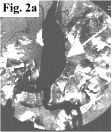
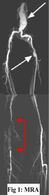
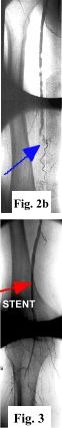
MRA FINDINGS: Fig. 1 on the left is the subtraction CE-MRA. Fig. 2 on the right displays selective x-ray angiogram images performed by Dr. Gadallah. There is a large fusiform aneurysm of the distal aorta and focal aneurysm in the left common femoral artery (white arrows), shown equally well on both MRA and x-ray angio (Fig. 2a). But the interesting finding is in the distal right superficial femoral artery (SFA) where there is 12 cm long segment occlusion seen on MRA (Fig. 1, red arrowheads) with reconstitution at the popliteal artery via collaterals. There is an apparent filling defect (arrows) within the SFA consistent with thrombosis. The surrounding hyperintensity probably represents enhancement in an edematous/inflamed vessel wall. The x-ray angio in Fig. 2b confirms total occlusion of distal SFA (blue arrows).
TREATMENT: The patient underwent angioplasty and stent placement by Dr. Gadallah with resultant restoration of brisk flow in the distal right SFA (red arrow in Fig. 3, which is a post-stent image).*
CLINICAL SIGNS OF ARTERIAL THROMBOSIS: Acute arterial thrombosis classically presents with the 5 P's: Pain, Pulselessness, Pallor, Paresthesia, and Perishing cold. However, more chronic thrombosis (such as in this case) gives rise to collateral vessels and may present with symptoms of vascular claudication.
Q. What is the accuracy of Contrast-Enhanced MRA run-off?
A. It is extremely accurate when performed properly, and it correlates well with x-ray angiography as seen here. Some patients may undergo surgery based on CE-MRA without the need for conventional angiography. 4D reconstruction allows visualization at multiple angles not possible with x-ray angiography. Moreover, MRA is non-invasive!
For more information, you may call myself at (661) 949-8111, Dr. Mary Toft at 945-0821, or Dr. Gadallah at 538-2222. If you have an interesting case, please contact me for a clinical-radiological case presentation.
Ray H. Hashemi, M.D., Ph.D.,
Director
*Clinical information provided by Dr. Toft and angiographic/therapeutic information by Dr. Gadallah. Dr. Toft is an internist together with Dr. Gary Grubb. Dr. Gadallah is an interventional cardiologist together with Dr. Choudhary.