 |
ADVANCED IMAGING CENTER PHYSICIAN NEWS |
April 14, 2003 |
PARATHYROID ADENOMA:
ULTRASOUND, MRI, AND RADIONUCLIDE SPECT CORRELATION
 |
 |
 |
 |
| Fig.1 | Fig.2 | Fig.3 | Fig.4 |
|
ADVANCED IMAGING CENTER PHYSICIAN NEWS |
April 14, 2003 |
|
|
|
|
| Fig.1 | Fig.2 | Fig.3 | Fig.4 |
CLINICAL INFORMATION: The patient is a 45-year-old female who presented to the office of Dr. Elea English* with high serum levels of calcium and parathyroid hormone PTH. Parathyroid adenoma was clinically suspected. She was referred to AIC for an ultrasound & MRI of the neck soft tissues and finally a nuclear Sestamibi parathyroid scan.
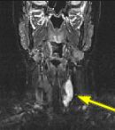
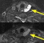
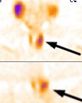
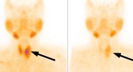
FINDINGS: The ultrasound (not shown) revealed a large solid mass behind the left lobe of the thyroid. Fig. 1-2 are MRI images of the neck demonstrating a mass behind the left thyroid lobe with hyperintensity on T2 weighted and STIR images (Fig. 1, 2 top) and enhancement higher than the thyroid (Fig. 2 bottom). Fig. 3-4 are Sestamibi images with Fig. 3 being coronal (top) and axial (bottom) SPECT images and Fig. 4 the early and delayed static images demonstrating persistent activity in the mass on delayed images while the thyroid activity washes out (arrows).
NORMAL SCAN: Normal physiologic uptake of Sestamibi can be seen within the salivary/parotid glands in addition to the thyroid as well as the heart. A normal parathyroid gland (and there are generally 4 of them: 2 superior and 2 inferior ones usually posterior to the upper and lower poles of the thyroid) does NOT show any uptake.
ABNORMAL SCAN: A parathyroid adenoma also demonstrates rapid uptake on the initial images. However, unlike a normal thyroid, the adenoma demonstrates persistent uptake of Sestamibi and is readily visible on delayed images. Ectopic adenomas in the mediastinum are also easily detected with this technique.
ACCURACY: Sestamibi scan is highly accurate in detecting parathyroid adenomas (sensitivity and specificity of about 95%). A combination of this scan and CT, MRI or US can detect and localize nearly 100% of adenomas.
CAUSES OF PRIMARY HYPERPARATHYROIDISM: Parathyroid Adenoma (87% = 80% single + 7% multiple); Parathyroid Hyperplasia (10%); Parathyroid Carcinoma (3%).
FINAL DIAGNOSIS: She underwent surgical resection of a parathyroid adenoma at UCLA and is now normo-calcemic.
For more information, you may call me at (661) 949-8111 or Dr. English at (661) 272-1400.
Ray H. Hashemi, M.D., Ph.D.
Director* Dr. Elea English, MD, is a family practitioner in Palmdale, California, and can be reached at (661) 272-1400.