 |
ADVANCED IMAGING CENTER PHYSICIAN NEWS |
February 7, 2000 |
MR Mammography (routine and contrast dynamic study): State-of-the-art
 |
 |
 |
 |
| |||
|
ADVANCED IMAGING CENTER PHYSICIAN NEWS |
February 7, 2000 |
|
|
|
|
| |||
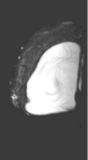
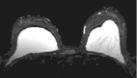
Q. What is MR Mammography and how is it performed?
MR Mammography is a fairly new MR procedure that evaluates breast parenchyma and implants. To be diagnostic, a high-resolution dedicated bilateral breast coil is required. The patient is placed in the prone position on the MR table. The breasts fall into the two cups of the dedicated breast coil. The patient is scanned in this position without changing her position throughout the study. Anything short of a high-resolution study is simply dangerous.
Q. What are the advantages of scanning in the prone position?
In the prone position, breasts are separated from the chest wall into a natural anatomical position thus eliminating artifacts from breathing and heart motion.
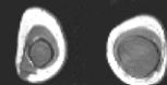
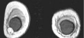
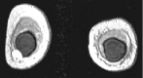
Q. What is the accuracy of MRI for diagnosing implant rupture?
High-resolution MRI is extremely accurate for diagnosing silicone and saline implant rupture. The so-called "linguini sign" refers to serpiginous thin lines seen with intracapsular rupture (left 3 images above). Various new "silicone techniques" can also detect extracapsular extravasation of silicone.
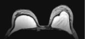
Q. What is the value of MRI for detection and diagnosis of breast cancer?
MRI is utilized in patients with dense breasts and indeterminate masses who have a non-diagnostic x-ray mammogram and ultrasound. MRI is only an adjunct technique. If a mass is suspected, a contrast-enhanced dynamic study is performed with IV Gadolinium. This is usually done with high-resolution (1-2 mm) coronal sections pre and post rapid infusion of contrast (using an MR-compatible power injector) in a dynamic fashion (usually 4 or 5 post contrast sequences are obtained as shown in top right 3 images). Contrast uptake graphs of suspicious masses are obtained. Malignancies tend to take up contrast more rapidly.
For more information, or for any questions or concerns, please call me personally.
Ray H. Hashemi, M.D., Ph.D.,
Director