 |
ADVANCED IMAGING CENTER PHYSICIAN NEWS |
February 1, 2003 |
TEMPORAL LOBE EPILEPSY
VALUE OF HIGH-RESOLUTION MR IMAGING OF THE HIPPOCAMPI
FOR MESIAL TEMPORAL SCLEROSIS AND HIPPOCAMPAL ATROPHY
 |
 |
 |
| Fig.1 | Fig.2 | Fig.3 |
|
ADVANCED IMAGING CENTER PHYSICIAN NEWS |
February 1, 2003 |
|
|
|
| Fig.1 | Fig.2 | Fig.3 |
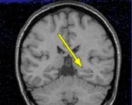
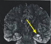
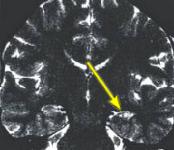
CLINICAL PRESENTATION: This young patient had a history of intractable complex partial seizures or temporal lobe epilepsy. He was referred for a high-resolution MRI of the brain and temporal lobes (hippocampi) in order to determine presence of mesial temporal sclerosis (MTS) and/or hippocampal atrophy. MRI is very important in planning for epilepsy surgery because of its ability to detect temporal lobe lesions that can cause seizures.
MRI FINDINGS: MRI detects mesial temporal sclerosis by demonstrating size asymmetry and abnormal signal within the atrophied hippocampus. Thin-section, high-resolution oblique coronal MR images are best for detecting these abnormalities, which can be subtle. Heavily T1-weighted images (Fig. 1) are best for detecting size asymmetry of the hippocampal gyri, while T2-weighted images (Fig. 3) and particularly FLAIR (fluid attenuated inversion recovery) images are most sensitive for detecting signal abnormalities (Fig. 2). The above images demonstrate left hippocampal atrophy and mesial temporal sclerosis (arrows).
ETIOLOGY: The most common cause of complex partial seizures is mesial temporal sclerosis (MTS), occurring in 35 to 65 percent of patients who undergo temporal lobe surgery. In mesial temporal sclerosis, the hippocampus is smaller than normal. This usually occurs on one side of the brain, but can occur bilaterally in 10 to 15 percent of cases. The cause of the neuronal loss is not known but may be related to repeated seizure activity (which places such high metabolic demands on neurons and eventually their destruction). Febrile seizures are also believed to be a potential cause of MTS.
Other Causes of Temporal Lobe Epilepsy: In addition to MTS, other temporal lobe lesions can cause seizures, including benign and malignant neoplasms, cortical dysplasia, vascular malformations and post-traumatic brain injury. MRI is again an excellent modality to evaluate all these possibilities. So if you have a patient with a history of seizures (especially temporal lobe epilepsy), consider MRI of the brain and hippocampi with/without contrast.
Temporal lobectomy can cure seizures in some patients with mesial temporal sclerosis. The patients who have the best outcomes after temporal lobectomy with mesial temporal sclerosis and temporal lobe epilepsy are those who have had pre-operative EEG showing seizure activity in the lobe that is removed, and patients whose epilepsy is not associated with generalized seizures.
Ray H. Hashemi, M.D., Ph.D.
Director