|
ADVANCED IMAGING CENTER PHYSICIAN NEWS |
September 18, 2000 |
INTERESTING CASE PRESENTATION:
RHEUMATOID ARTHRITIS
|
ADVANCED IMAGING CENTER PHYSICIAN NEWS |
September 18, 2000 |
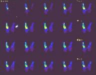
Fig. 1a
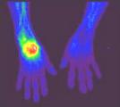
Fig. 1b
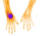
Fig. 1c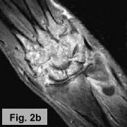
CLINICAL PRESENTATION: This 60-year-old female patient presented to the office of Dr. Sethu Madhavan and Dr. Nitin Shah with pain and swelling in the left wrist. Initially, cellulitis was suspected and the patient was referred to AIC for a triple-phase bone scan in order to rule out osteomyelitis. Subsequently, an MRI of the left wrist was also requested to narrow down the differential diagnosis.*
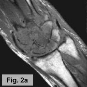
IMAGING FINDINGS: A triple-phase bone scan was obtained on AIC's dual-head nuclear camera. Fig. 1a-c show the angiographic (flow) images, blood pool images, and delayed (static) images of the wrists, respectively. They demonstrate marked increased uptake in the left wrist in all 3 phases of the study. Fig. 2a-b are selective pre and post-contrast MRI images of the left wrist showing precontrast T1-weighted (Fig. 1a) and postcontrast T1-weighted with Fat SAT (Fig. 1b). They demonstrate diffuse edema in the marrow of all the carpal bones and proximal metacarpal bones with diffuse enhancement of the marrow and surrounding synovium. X-rays (not shown) revealed diffuse osteopenia and erosions of the carpal bones.
DIFFERENTIAL DIAGNOSIS: The bone scan findings are usually seen with osteomyelitis, but diffuse carpal involvement is unusual. Increased uptake on all 3 phases can also be seen with Reflex Sympathetic Dystrophy (RSD) or an inflammatory process. The MRI and x-ray findings are more compatible with an inflammatory process, particularly rheumatoid arthritis, with diffuse synovitis and erosions and reactive marrow changes, although this is a late presentation for the patient's age.
FINAL DIAGNOSIS: Further lab results revealed marked elevation of rheumatoid factor. The diagnosis is, therefore, aggressive rheumatoid arthritis. Surgical treatment includes synovectomy.
For more information, you may call me at (661) 949-8111, Dr. Shah at (661) 945-7802, or Dr. Madhavan at 945-7181.
Ray H. Hashemi, M.D., Ph.D.
Director
*Clinical information provided by Dr. Nitin Shah, MD, an orthopedic surgeon in Lancaster, and Dr. Sethu Madhavan, an internist and gastroenterologist in Lancaster.