 |
ADVANCED IMAGING CENTER PHYSICIAN NEWS |
August 14, 2000 |
Advanced Topics in Neuroradiology:
Value of MRI FLAIR Sequence in Multiple Sclerosis
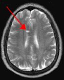 Fig. 1 |
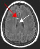 Fig. 2 |
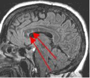 Fig. 3 |
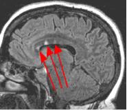 Fig. 4 |
|
ADVANCED IMAGING CENTER PHYSICIAN NEWS |
August 14, 2000 |
Fig. 1 |
Fig. 2 |
Fig. 3 |
Fig. 4 |
Q. What is a FLAIR MRI sequence?
FLAIR stands for FLuid Attenuated Inversion Recovery. It is a special MRI pulse sequence in which fluid such as CSF appears dark and most lesions including MS plaques, other white matter lesions, tumors, edema, and acute infarcts appear bright.
Q. What are the advantages of FLAIR?
By turning CSF dark, bright periventricular lesions such as MS plaques become much more conspicuous. Fig. 1 shows a typical T2-weighted axial image of the brain in which CSF in the ventricles appears bright. A lesion on the right is present (red arrow) that is also bright similar to CSF. Fig. 2 is a corresponding FLAIR image showing an obvious bright MS plaque (white arrow) against a background of dark CSF and gray parenchyma with additional more subtle lesions.
Q. What are the advantages of performing thin-section sagittal FLAIR?
Thin-section sagittal FLAIR images increase detection of small MS plaques even further, first reported by Hashemi, et al. (Ref. 1). They also have the advantage of clearly showing the corpus callosum and subependymal regions. An early sign of MS is subependymal nodularity and subcallosal striations (Ref. 1-2), not clearly visible on axial images. Fig. 3-4 in the same patient clearly show subcallosal MS plaques (arrows) along the undersurface of corpus callosum.
Axial FLAIR sequence is a part of our routine brain protocol at AIC. Thin-section sagittal FLAIR is also performed as an additional sequence when MS is a suspected diagnosis. For more information, you may call me at (661) 949-8111.
Ray H. Hashemi, M.D., Ph.D.,
DirectorREFERENCES:
- Ray H. Hashemi, William G. Bradley, et al. "Suspected Multiple Sclerosis: MR Imaging with a Thin-Section Fast FLAIR Pulse Sequence." Radiology 1995 Aug; 196(2):505-510.
- Palmer S, Bradley WG, et al., "Subcallosal striations: early findings of multiple sclerosis on sagittal, thin-section, fast FLAIR MR images." Radiology 1999 Jan; 210(1):149-153.