|
ADVANCED IMAGING CENTER PHYSICIAN NEWS |
July 10, 2000 |
INTERESTING CASE PRESENTATION
High-resolution MRI of the HAND with pathologic correlation
|
ADVANCED IMAGING CENTER PHYSICIAN NEWS |
July 10, 2000 |
CLINICAL PRESENTATION: This 34-year-old male patient presented to the office of Dr. A. Rahmati with a small lump and pain in the volar aspect of the right hand about the 4th and 5th fingers with stiffness and some numbness. Patient was referred to AIC for a high resolution MRI of the hand. *
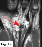
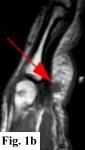
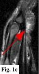
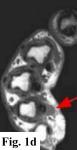
MRI FINDINGS: MRI was performed on AIC's high-field 1.5 Tesla short-bore Siemens Symphony scanner using a small flexible surface coil. Fig. 1a is a high-resolution coronal DESS, Fig. 1b a sagittal T1 weighted, Fig. 1c a sagittal turbo STIR, and Fig. 1d an axial T1 weighted. They demonstrate a 5x5x7 mm nodule in the volar aspect of the hand at the level of the 4th MCP joint inseparable from the flexor digitorum superficialis tendon (arrows) with some adjacent soft tissue edema. The underlying tendon and bones are otherwise intact.
DIFFERENTIAL DIAGNOSIS: Based on the MRI findings the DDX includes a benign soft tissue tumor such as a xanthoma, fibroma, hemangioma, neuroma or a lesion related to the tendon sheath such as a giant cell tumor of the tendon sheath or tendinous xanthoma. An inflammatory node was also in the differential. It does not have the MRI characteristics of a cyst or ganglion.
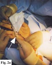
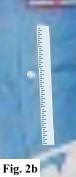
SURGICAL-PATHOLOGIC DIAGNOSIS: The patient was taken to surgery at LCH by Dr. Rahmati and a "rather solid round mass attached to the volar aspect of the 4th tendon sheath" (arrows in Fig. 2a-b) was excised measuring about 4x4x3 mm. Pathology at LCH revealed a "grayish-white somewhat nodular smooth soft tissue measuring 9x8x5 mm" with histopathologic "features in favor of a giant cell tumor of tendon sheath" interpreted as benign.
Q. What is a Giant Cell Tumor of Tendon Sheath?
It belongs to xanthomatoses, which consist of a group of tumor-like proliferations characterized by the presence of a variable number of foam cells. Tendinous xanthomas are common about the fingers, heel, elbow and knee, where they may erode the bone. A giant cell tumor of the tendon sheath is detected in the hands and feet, attached to tendons, tendon sheaths, of fibrous capsules. They are benign but may recur following resection.
For more information, you may call myself at (661) 949-8111, or Dr. Rahmati at 723-3434. If you have an interesting case, please contact me for a clinical-radiological case presentation.