 |
ADVANCED IMAGING CENTER PHYSICIAN NEWS |
July 2, 2001 |
INTERESTING CASE PRESENTATION
Slipped Capital Femoral Epiphysis (SCFE): Early MRI Findings
 |
 |
 |
 |
|
ADVANCED IMAGING CENTER PHYSICIAN NEWS |
July 2, 2001 |
|
|
|
|
CLINICAL INFORMATION: The patient is a 10-year-old female who presented with right hip pain to Dr. Satey*. The x-rays (not shown) presumably were equivocal for growth plate widening but otherwise unremarkable. The patient was referred to AIC for an MRI of the hips.
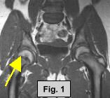
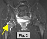
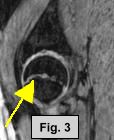
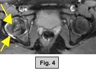
MRI FINDINGS: The MRI was performed on AIC's high-field 1.5 Tesla short-bore Siemens Symphony. Coronal T1 and fat suppressed turbo STIR weighted, axial proton density and T2 weighted with Fat Saturation, and sagittal DESS sequences were obtained bilaterally. Fig. 1 (coronal T1W) shows widening and irregularity of the growth plate in the right hip. Fig. 2 (coronal STIR) shows subtle adjacent marrow edema and moderate joint effusion. Fig. 3 (sagittal DESS) and Fig. 4 (axial PD Fat Sat) also demonstrate the same findings. Subtle medial displacement of the right femoral epiphysis is also noted on the coronal images.
DIAGNOSIS: The above findings are consistent with Slipped Capital Femoral Epiphysis (SCFE).
CAUSES OF HIP PAIN IN A CHILD: They include SCFE, avascular necrosis (Legg-Calve-Perthes disease), septic arthritis, fracture, dislocation, transient osteoporosis, etc. The diagnosis depends on the age, clinical presentation and/or radiographic findings.
DISCUSSION: SCFE is basically a Salter-Harris type I injury to the growth plate. It usually occurs in obese adolescent males but can occur in females as well. About 10% of the cases are bilateral. SCFE is usually a sequela of chronic stress/trauma where subclinical stresses are applied to the growth plate, and slowly there is slippage medially and posteriorly (best seen on frog leg views). Eventually clear-cut slippage of the femoral head will occur.
ETIOLOGY: Chronic stress/trauma, or weakened bones such as due to renal osteodystrophy, rickets, childhood irradiation, hypothyroidism, growth hormone deficiency, etc.
TREATMENT: Treatment is surgical. This patient was referred to Dr. Sobeck*. Diagnosis of SCFE was confirmed and the patient underwent percutaneous pinning of the right femoral physis.
For more information, please call Dr. Satey, Dr. Sobeck or myself at the numbers below.
Ray H. Hashemi, M.D., Ph.D.
Director* Dr. Satey, MD, is a pediatrician in Palmdale, California, and can be reached at (661) 272-5656.
* Dr. Sobeck, MD, is an orthopedic surgeon in Lancaster and Sherman Oaks and can be reached at (661) 940-9606.