 |
ADVANCED IMAGING CENTER PHYSICIAN NEWS |
May 14, 2002 |
INTERESTING CASE PRESENTATION
Obstructive Tumor of Aqueduct
|
ADVANCED IMAGING CENTER PHYSICIAN NEWS |
May 14, 2002 |
CLINICAL PRESENTATION: This patient in his early forties presented to the office of Dr. A. Farrukh and Dr. M.K. Dhillon* with progressive intermittent headaches and some balance and bladder dysfunction. An outside scan (not shown) suggested possible obstruction at the level of the aqueduct of Sylvius. The patient was referred to AIC for high-resolution MRI of the brain with 1 mm sections through the aqueduct and CSF flow study.
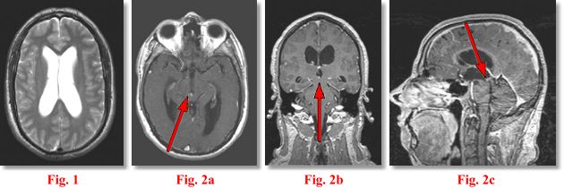
IMAGING FINDINGS: The study was performed on AIC’s high-field Siemens Symphony scanner. Fig. 1 shows an axial T2 weighted image of the ventricles demonstrating moderate hydrocephalus. Fig. 2a-c are post-contrast axial (5 mm), coronal (1 mm) and sagittal (1 mm) T1 weighted images through the aqueduct. They demonstrate a subtle tiny 4 mm enhancing mass (arrows) at the entry zone into the aqueduct at the junction with the inferior 3rd ventricle.
DIFFERENTIAL DIAGNOSIS: This is a solid mass in the ventricular system. The following DDX was rendered at the time of the scan: meningioma, ependymoma, subependymoma, PNET, a vascular lesion or, less likely, a glioma or metastasis.
SURGICAL PROCEDURE: The patient underwent surgery at UCLA Medical Center by Neurosurgeon Dr. Marvin Bergsneider including endoscopic ventriculocisternostomy and endoscopic-assisted tumor biopsy and excision. A recent follow-up MRI done at AIC (not shown) showed complete removal of the tumor and resolution of hydrocephalus.
PATHOLOGIC DIAGNOSIS: Pathology revealed a PINEOCYTOMA, obviously in an ectopic location as the tumor was away from the pineal gland, which is posterior to the 3rd ventricle. Final Diagnosis: Ectopic Pineocytoma.
This was probably the smallest obstructive intracranial tumor I have ever seen!!! For more information, you may call me at (661) 949-8111 or Drs. Farrukh and Dhillon at (661) 945-6931.
Ray Hashemi, MD, PhD
Director