 |
ADVANCED IMAGING CENTER PHYSICIAN NEWS |
March 26, 2001 |
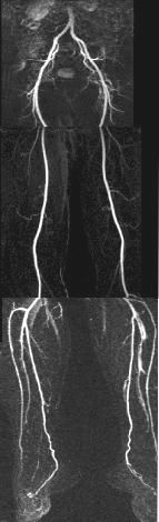
We are pleased to announce the introduction of an automatic stepping-table technology to our MRA run-off studies on the AIC's high-field 1.5 Tesla short-bore Siemens Symphony MR scanner. AIC is the only site in the Antelope Valley with this revolutionary technology. In fact, there are only a few sites in the LA area with this capability. What are the advantages of a moving table? The MRA run-off covers the distal aorta to the arteries in the feet. The stepping table feature automatically advances the MR table so that the areas of interest are fully covered in 3 successive sections. This allows accurate pre and post contrast and delayed studies in exact identical sections for subtraction purposes. It also removes errors due to manual advancement of the table. This technology is possible through addition of a new computer software contolling a new stepping table hardware. TECHNIQUE: Contrast-enhanced MR Angiography (CE-MRA) at AIC is performed utilizing ultrafast coronal sections during intravenous infusion of 30-40 cc of gadolinium contrast using an MR-compatible power injector at a fast programmed rate. Subsequently, subtraction images and 4D reconstructions are obtained. Each section is obtained in about 30 seconds using state-of-the-art ultrafast MR techniques. The stepping table advances automatically twice during each run. Two back-to-back runs are obtained. Subtraction is performed by subtracting the pre-contrast from post-contrast images. EXAMPLE: The example shown here is on our volunteer Dr. Udkoff, who is fortunately asymptomatic and demonstrates a normal scan. As you can see, the aorta and iliac, femoral, and popliteal arteries and trifurcations through the calf arteries all the way into the feet are exquisitely depicted. These images are comparable to image quality of conventional x-ray angiography. In fact, the calf arteries are generally depicted better than x-ray angiography. CONCLUSION: Compared to conventional x-ray angiography, MRA run-off is a relatively non-invasive examination of the aorta and extremities (thanks to intravenous injection instead of transarterial catheterization, and use of gadolinium instead of iodinated contrast). With the introduction of stepping-table technology, MRA run-off is comparable to, if not better than, conventional angiography in terms of ease and diagnostic capabilities. The beauty of MRA is that 4D images can be rotated in every which way, a feature not possible with conventional angiography in which additional views require additional injection of iodine contrast, a potential hazard to the kidneys. MRI contrast, gadolinium, is on the other hand very safe and only a total of 30-40 cc is injected. We propose that with the availability of current moving stepping-table technology and ultrafast MR techniques, MRA run-off should be the first modality of choice for all run-off studies, unless there is a contraindication to MRI such as pacemakers. Eventually, vascular surgeons will learn to appreciate the tremendous value of this new technology and will shift their preference from invasive x-ray angiography to non-invasive MRA run-off angiography. Needless to say, contrast-enhanced MRA is equally valuable for evaluation of carotid and renal arteries.
Ray H. Hashemi, M.D., Ph.D. |
| Home Page | Procedures | Certification | Radiologists | News Releases | FAQs | About Us | Location | Contact Us | 3D Gallery | Glossary | Frames |
ADVANCED IMAGING CENTER
43731 N. 15th Street West, Lancaster, CA 93534
Phone (661) 949-8111 - Fax (661) 949-6600