|
ADVANCED IMAGING CENTER PHYSICIAN NEWS |
February 26, 2001 |
Ultrafast Contrast-Enhanced Dynamic MRI of the Pituitary Gland
State-of-the-art Imaging of Pituitary Microadenomas
|
ADVANCED IMAGING CENTER PHYSICIAN NEWS |
February 26, 2001 |
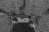
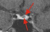
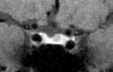
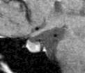
Fig. 1 Fig. 2 Fig. 3a Fig. 3b CLINICAL PRESENTATION: This young female was referred to AIC for diagnosis of a possible pituitary microadenoma (prolactinoma). A contrast-enhanced (CE) dynamic MRI scan was performed.
MRI TECHNIQUE: The scan is done on AIC's high-field 1.5 Tesla Siemens Symphony short-bore MR scanner. Following routine scan of the brain and sella, a sequential ultrafast coronal scan of the pituitary gland is set up. Each scan is performed in about 15-20 seconds, and multiple back-to-back serial scans are obtained during rapid intravenous infusion of 10-20 cc of gadolinium contrast using an MR-compatible power injector. Delayed scans in the coronal and sagittal planes are also obtained through the sella.
MRI FINDINGS: Fig. 1 is a precontrast coronal image through the sella. Fig. 2 is an image from the first postcontrast dynamic scan. Fig. 3a-b are delayed coronal and sagittal scans. A 6 mm microadenoma is clearly visible in Fig. 2 in the left aspect of the gland (red arrows). This is due to the fact that normal pituitary tissue (owing to lack of blood brain barrier) rapidly enhances before a microadenoma demonstrates enhancement. Note that this same lesion is less conspicuous on the delayed scans (Fig. 3a-b). In some cases, delayed scans completely obscure a microadenoma due to the fact that the microadenoma gradually enhances as time goes by. Routine MRI of the sella utilizes scans that are on the order of minutes, allowing a microadenoma to enhance thus reducing tissue contrast between a microadenoma and normal pituitary tissue.
CONCLUSION: Dynamic MRI of the sella should be the modality of choice for detection of pituitary microadenomas including prolactinomas.
REFERENCE: The following is just one pertinent reference:
Corticotropin- and thyrotropin-secreting pituitary microadenomas: detection by dynamic magnetic resonance imaging. Smallridge RC; et al., Mayo Clin Proc 2000 May;75(5):521-8. The authors conclude: "We recommend that dynamic MRI be performed in any patient with a suspected microadenoma, before IPSS (surgical sampling) is performed."
For more information, you may call me at (661) 949-8111.
Ray Hashemi, MD, PhD
Director