Since their discovery in 1895, x-rays have been a vital
scientific tool, revealing previously concealed worlds. Because of their
great penetrating power, x-rays can also be used to study the structure of
living organisms.
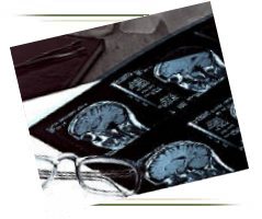
One of the earliest applications of x-rays was in
medicine, where they were used for both diagnosis and therapy. They
penetrate soft tissues but are stopped by bones, which absorb them. Thus
if a photographic plate that is sensitive to x-rays is placed behind a
part of the body and an x-ray source is placed in front, x-ray exposure
will result in an image of the bones and internal organs. When the
radiograph, or plate, is developed, a negative image is produced.
Tissues that are easily penetrated by x-rays appear dark, while bones and
dense tissues show up as light or white regions.
Although bones are the most opaque structures, there
are many dense tissues, such as tumors, that can also show up unusually
light in radiographs. These images can be used to study damaged or
broken bones, inspect dental cavities, detect foreign objects in the body,
and diagnose diseases.
To utilize x-rays for the investigation of other, less
dense tissues of the body, such as the gastrointestinal tract, the tissues
must first be made opaque to x-rays. Generally, patients are asked to
drink a mixture containing an opaque substance, such as barium, so that
the outline of the digestive tract becomes visible with x-rays.
The clinic also offers fluoroscopy, a similar technique
that uses x-rays to observe a particular organ in action, such as the
heart. The patient is placed in the fluoroscope between the x-ray source
and a screen that is coated with a fluorescent substance. The image that
appears when the x-rays strike the screen is brightened by an electronic
device called an image intensifier.
| ScanHealth |
Open MRI |
High-field MRI | MR Angiography |
Helical CT |
CT Angiography | Calcium Scoring
|
| 4D CT Reconstruction |
Dental Scan | 4D
Ultrasound | Nuclear Medicine |
PET Scan |
DEXA Bone Density | X-ray |
 1.
The only community-based, private-practice, physician-operated
imaging facility in the Antelope Valley, just like any other private
practice medical office. Not belonging to any hospital or outside
imaging network. This means more personal and caring service.
1.
The only community-based, private-practice, physician-operated
imaging facility in the Antelope Valley, just like any other private
practice medical office. Not belonging to any hospital or outside
imaging network. This means more personal and caring service.